Back Witstof Afrikaans المادة البيضاء Arabic Ağ maddə Azerbaijani Bijela masa BS Substància blanca Catalan Bílá hmota Czech Hvid substans Danish Weiße Substanz German Λευκή ουσία Greek Blanka substanco Esperanto
| White matter | |
|---|---|
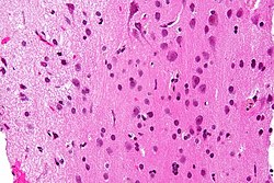
 Micrograph showing white matter with its characteristic fine meshwork-like appearance (left of image – lighter shade of pink) and grey matter, with the characteristic neuronal cell bodies (right of image – dark shade of pink). HPS stain. | |
 Human brain right dissected lateral view (anterior on the right), showing grey matter (the darker outer parts), and white matter (the inner and prominently whiter parts). | |
| Details | |
| Location | Central nervous system |
| Identifiers | |
| Latin | substantia alba |
| MeSH | D066127 |
| TA98 | A14.1.00.009 A14.1.02.024 A14.1.02.201 A14.1.04.101 A14.1.05.102 A14.1.05.302 A14.1.06.201 |
| TA2 | 5366 |
| FMA | 83929 |
| Anatomical terminology | |

White matter refers to areas of the central nervous system (CNS) that are mainly made up of myelinated axons, also called tracts.[1] Long thought to be passive tissue, white matter affects learning and brain functions, modulating the distribution of action potentials, acting as a relay and coordinating communication between different brain regions.[2]
White matter is named for its relatively light appearance resulting from the lipid content of myelin. However, the tissue of the freshly cut brain appears pinkish-white to the naked eye because myelin is composed largely of lipid tissue veined with capillaries. Its white color in prepared specimens is due to its usual preservation in formaldehyde.
- ^ Blumenfeld, Hal (2010). Neuroanatomy through clinical cases (2nd ed.). Sunderland, Mass.: Sinauer Associates. p. 21. ISBN 978-0878936137.
Areas of the CNS made up mainly of myelinated axons are called white matter.
- ^ Douglas Fields, R. (2008). "White Matter Matters". Scientific American. 298 (3): 54–61. Bibcode:2008SciAm.298c..54D. doi:10.1038/scientificamerican0308-54.